
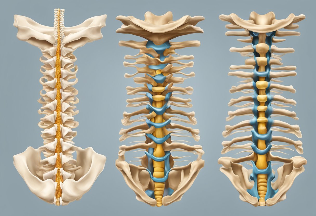
The cervical spine, also known as the neck, is a crucial part of the human body that supports the weight of the head and allows for its movement. This section of the spine consists of seven vertebrae, labeled C1 through C7. Each of these vertebrae has its own unique structure and function, which work together to provide flexibility and stability to the neck.
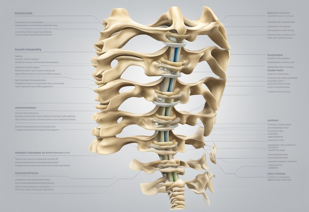
The first two vertebrae of the cervical spine, C1 and C2, are particularly important as they support the weight of the head and allow for its rotation. C1, also known as the atlas, is shaped like a ring and sits directly beneath the skull. It is responsible for supporting the weight of the head and allowing for its nodding motion. C2, also known as the axis, has a unique structure that allows for the rotation of the head. Its most distinctive feature is the dens, a bony projection that protrudes upward and fits into a space in the atlas.
The remaining five vertebrae of the cervical spine, C3 through C7, are responsible for providing flexibility and stability to the neck. These vertebrae are larger and more robust than the upper two, and are designed to withstand greater forces. They are connected by intervertebral discs, which act as shock absorbers and allow for movement between the vertebrae. Understanding the components of the cervical spine is crucial for diagnosing and treating neck injuries and conditions, and for maintaining overall spinal health.
Anatomy of the Cervical Spine
The cervical spine, or neck, is composed of seven vertebral bodies stacked on top of each other. Each vertebral body has a hole in the center, through which the spinal cord passes. The cervical spine is responsible for supporting the weight of the head and allowing for movement in multiple directions.
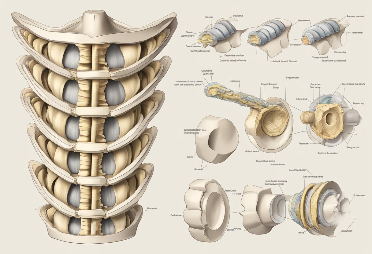
Vertebral Bodies
The vertebral bodies of the cervical spine are small and delicate, with a characteristic oval shape. They are separated by intervertebral discs, which act as shock absorbers and allow for movement between the vertebrae. The vertebral bodies are also connected by facet joints, which allow for smooth gliding movements between the vertebrae.
Intervertebral Discs
The intervertebral discs of the cervical spine are made up of a tough outer layer called the annulus fibrosus and a gel-like center called the nucleus pulposus. These discs are responsible for absorbing shock and distributing pressure evenly across the cervical spine. Over time, the intervertebral discs can degenerate, leading to conditions such as herniated discs and spinal stenosis.
Facet Joints
The facet joints of the cervical spine are located on the back of the vertebrae and allow for smooth gliding movements between the vertebrae. These joints are lined with cartilage and surrounded by a capsule filled with synovial fluid, which lubricates the joint and reduces friction. The facet joints are also responsible for limiting excessive movement in the cervical spine.
In summary, the cervical spine is composed of seven small and delicate vertebral bodies, separated by intervertebral discs that act as shock absorbers and allow for movement between the vertebrae. The facet joints allow for smooth gliding movements between the vertebrae and limit excessive movement in the cervical spine.
Cervical Spine Function
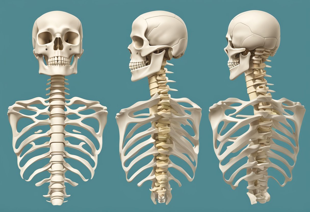
The cervical spine, also known as the neck, is made up of seven vertebrae that provide support and mobility to the head and neck. It plays a crucial role in the overall function of the spine and body.
Support and Structure
The cervical spine provides support and structure to the head and neck, which allows for proper alignment and balance of the body. The vertebrae in the cervical spine are smaller than those in the thoracic and lumbar regions, but they have a unique shape that enables them to support the weight of the head and allow for movement.
The cervical spine also houses and protects the spinal cord, which is the main pathway for communication between the brain and the rest of the body. The spinal cord runs through the center of the vertebrae and is surrounded by protective layers of bone and soft tissue.
Range of Motion
The cervical spine has a wide range of motion, which allows for movement of the head and neck in various directions. This range of motion is due to the unique shape of the cervical vertebrae, which allows for flexion, extension, rotation, and lateral bending.
The muscles, ligaments, and tendons surrounding the cervical spine provide additional support and stability during movement. These structures work together to allow for smooth and controlled movement of the head and neck.
In summary, the cervical spine plays a critical role in providing support and structure to the head and neck while allowing for a wide range of motion. Understanding the function of the cervical spine is essential for maintaining proper alignment and balance of the body.
Nervous System Components
The cervical spine is responsible for protecting the vital nervous system components that control the body’s functions and movements. The nervous system components of the cervical spine include the spinal cord and nerve roots.
Spinal Cord
The spinal cord is a long, thin, tubular bundle of nervous tissue that extends from the brainstem to the lumbar region of the spine. It is protected by the bony structures of the spine and is responsible for transmitting signals between the brain and the rest of the body. The spinal cord is divided into segments, each of which corresponds to a specific region of the body.
Nerve Roots
The nerve roots are the points at which the spinal cord branches out into the peripheral nervous system. The cervical spine has eight pairs of nerve roots, each of which corresponds to a specific region of the body. These nerve roots exit the spine through small openings between the vertebrae and then branch out to innervate the various muscles, organs, and tissues of the body.
In summary, the nervous system components of the cervical spine play a crucial role in controlling the body’s functions and movements. The spinal cord and nerve roots work together to transmit signals between the brain and the rest of the body, allowing us to move, feel, and interact with our environment.
Ligaments and Muscles
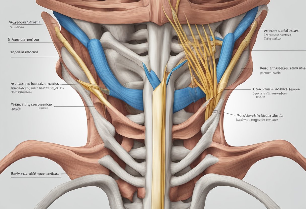
Anterior and Posterior Ligaments
The cervical spine is held together by a network of ligaments that provide stability and support. The anterior longitudinal ligament (ALL) runs along the front of the vertebral bodies, while the posterior longitudinal ligament (PLL) runs along the back. The ALL prevents excessive extension of the spine, while the PLL prevents excessive flexion.
In addition to the ALL and PLL, there are several other important ligaments in the cervical spine. The ligamentum flavum connects the laminae of adjacent vertebrae, while the interspinous and supraspinous ligaments connect the spinous processes. These ligaments help to limit excessive movement between the vertebrae and provide additional support to the spine.
Musculature
The muscles of the cervical spine are responsible for movement and stability. The deep neck flexors, including the longus colli and longus capitis muscles, are responsible for flexion of the neck. The sternocleidomastoid muscle, which runs from the sternum and clavicle to the mastoid process of the temporal bone, is responsible for rotation and lateral flexion of the neck.
The trapezius muscle, which runs from the occipital bone to the thoracic spine and shoulder blade, is responsible for elevation and retraction of the scapula. The levator scapulae muscle, which runs from the cervical spine to the scapula, is responsible for elevation of the scapula. The scalene muscles, which run from the cervical spine to the first and second ribs, are responsible for elevation of the ribs during inspiration.
Overall, the ligaments and muscles of the cervical spine work together to provide stability and support while allowing for a wide range of movement.
Vascular Elements
The cervical spine contains several vital vascular elements that supply blood to the brain and spinal cord. These elements include the vertebral arteries and venous plexuses.
Vertebral Arteries
The vertebral arteries are two small arteries that originate from the subclavian arteries and enter the cervical spine through the transverse foramina of the cervical vertebrae. They then join together to form the basilar artery at the base of the skull. The vertebral arteries provide blood supply to the posterior part of the brain, including the brainstem and cerebellum.
Venous Plexuses
The venous plexuses in the cervical spine are a network of veins that drain blood from the spinal cord and surrounding tissues. There are three main venous plexuses in the cervical spine: the anterior, middle, and posterior plexuses. The anterior and middle plexuses drain into the vertebral veins, while the posterior plexus drains into the vertebral venous plexus. The vertebral venous plexus then drains into the brachiocephalic veins, which eventually empty into the superior vena cava.
Overall, the vascular elements of the cervical spine play a crucial role in supplying blood to the brain and spinal cord. Understanding the anatomy and function of these elements is important for diagnosing and treating cervical spine disorders.